
Panoramic Dental X Ray: Understanding the Process
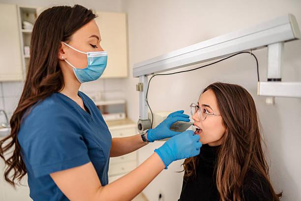
A Panoramic Dental X Ray in Dubai is a dental imaging procedure that captures a broad view of the teeth, upper and lower jaws, and surrounding oral structures in one image. Unlike small intraoral X-rays that focus on particular teeth, panoramic imaging provides a wider perspective, making it useful for different types of dental assessments.
The process is generally quick and non-invasive. Patients are positioned carefully in front of a specialized X-ray machine, which rotates around the head while capturing the necessary images. These images are combined to produce a panoramic view that a dental professional can evaluate.
Panoramic imaging may be recommended for wisdom tooth assessment, orthodontic planning, evaluation of developing teeth, missing teeth, or certain dental procedures. The dentist decides whether this type of X-ray is appropriate based on the patient's individual needs.
Preparing Before the Scan
Preparation for a panoramic X-ray is usually simple. Before the procedure, the dental professional may ask you to remove items that could interfere with the image. These may include earrings, necklaces, glasses, or certain removable accessories around the head and neck.
You should also inform the dental team if you are pregnant or think you may be pregnant. Providing accurate information about your dental history and previous imaging can also help the dental professional determine the most suitable approach.
There is generally no special fasting or complicated preparation required. Following the instructions provided before the scan is usually enough to ensure a smooth experience.
Dental Clinic: Positioning for the Panoramic Scan
At a Dental Clinic, proper positioning is an important part of the imaging process. The dental professional may ask you to stand or sit in front of the machine and place your chin on a designated support.
Depending on the equipment, you may also be asked to gently bite onto a positioning guide. This helps keep the teeth and jaws in the correct relationship while the machine captures the image.
Your head may be supported or stabilized to reduce movement. Remaining still is particularly important because movement during the scan can affect image quality and may require the image to be captured again.
How the Machine Captures the Image
Once you are positioned correctly, the panoramic machine begins to move around your head. The equipment captures information from different angles as it rotates.
The system then combines this information into a single panoramic image. This image provides a broad view of the mouth and jaw rather than focusing on just one tooth.
The actual scanning process usually takes only a short time. Patients do not normally feel anything while the image is being captured, making the procedure straightforward for most people.
What Does the Dentist Examine?
After the scan is complete, the dentist reviews the panoramic image. The image can provide information about the position of teeth, developing teeth, impacted wisdom teeth, missing teeth, and general jaw structures.
The dentist may also look for areas that appear unusual and may require additional investigation. If more detail is needed, another type of dental X-ray or imaging examination may be recommended.
A panoramic image is therefore considered together with the patient's symptoms, dental history, and physical examination. It is not generally used as the only source of information when making a dental assessment.
Panoramic Imaging for Wisdom Teeth
Wisdom teeth are often assessed using panoramic imaging because they develop at the back of the mouth and may be difficult to evaluate visually.
A panoramic image can show the general position and development of wisdom teeth. It may help the dentist determine whether they are erupting normally or appear impacted.
If a wisdom tooth is angled or positioned unusually, the dentist can assess the image and decide whether further examination is needed. This can be especially useful when a patient has discomfort or other symptoms around the back teeth.
The Role of Panoramic X Rays in Orthodontics
Panoramic imaging can also be useful during orthodontic assessments. Dentists and orthodontic professionals may need to understand the position of multiple teeth before planning braces or other alignment options.
The image can show developing teeth, missing teeth, crowded areas, and general tooth positioning. This information may be combined with photographs, clinical examinations, and other diagnostic records to develop a more complete understanding of the patient's dental condition.
What Happens After the Scan?
Once the image has been captured and reviewed, the dentist explains any relevant findings. If everything that needs to be assessed is visible, no additional imaging may be necessary.
If a specific tooth or area requires greater detail, the dentist may recommend a targeted X-ray or another imaging technique. This depends on the reason for the original scan and what the dental professional observes.
There is normally no recovery period after a panoramic X-ray. Most patients can return to their usual activities immediately after the procedure.
Is Panoramic Imaging Safe?
Dental X-rays use controlled amounts of radiation to produce diagnostic images. Modern dental imaging systems are designed to minimize exposure while obtaining the information required for assessment.
Patients should always discuss pregnancy or possible pregnancy with their dental professional before undergoing an X-ray. The dentist can then consider the individual circumstances and determine whether imaging is appropriate.
Why Following Instructions Matters
Proper positioning and remaining still are important parts of obtaining a clear panoramic image. A blurred or incorrectly positioned image may make it harder for the dentist to evaluate the structures accurately.
Simple instructions such as keeping your head still, maintaining the required bite position, and removing interfering objects can help improve the quality of the scan.
Final Thoughts
Understanding the panoramic imaging process can help patients feel more prepared before their dental scan. From careful positioning to image capture and professional interpretation, each step contributes to obtaining a useful view of the teeth and jaw. When appropriately recommended, Panoramic Dental X Ray imaging can provide valuable information and support a clearer assessment of overall oral health.

 English
 Arabic
 Spanish
 Portuguese
 Deutsch
 Turkish
 Dutch
Dutch
 Italiano
 Russian
 Romaian
 Portuguese (Brazil)
 Greek
Greek